Tooth #13 (#6) presents in a cross-bite position
Depp probing depths were noted along with class III mobility.
A CBCT Scan confirmed the clinical findings and rendered a hopeless prognosis for the tooth
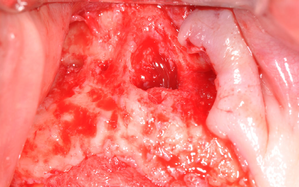
Upon extraction and flap reflection a fenestration was noted at the apical aspect of the buccal plate.
Complete loss of the palatal plate was noted
The site was augmented with a combination of Freeze-Dried bone allograft and bovine xenograft. An absorbable collagen membrane (Bio-gide) was used to cover the buccal fenestration, and a non-absorbable d-PTFE Ti reinforced membrane was used to resemble the missing palatal wall.
The buccal flap was advanced to cover the membrane, however, primary closure was not achieved.
Exposure of the membrane was noted after two weeks of healing.
No signs of infection were noted and the membrane was removed 8 weeks following the augmentation of the deficient site.
Complete epithelialization of the site was noted 2 weeks after the membrane was removed
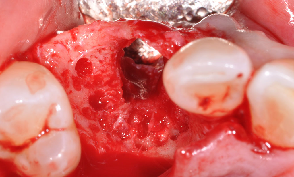
Complete reconstruction of the ridge was noted 4 months following the augmentation procedure.
A CBCT Scan revealed adequate bone volume for implant placement
Clinical measurement revealed a 7-8mm wide ridge with no vertical deficiency
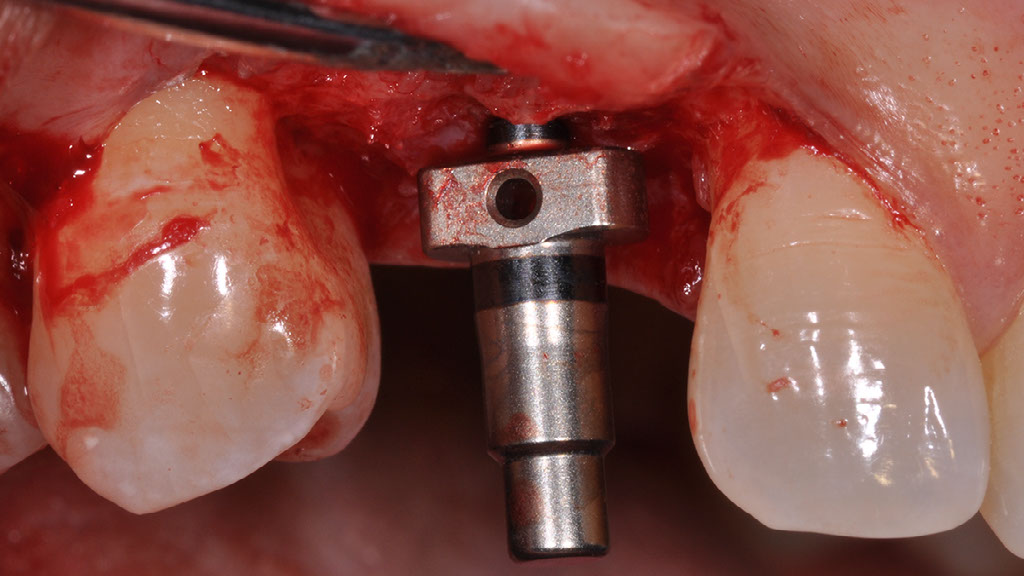
An Astra, Osseospeed EV implant, 4.2x9.0mm was placed, with insertion torque of >35N/cm
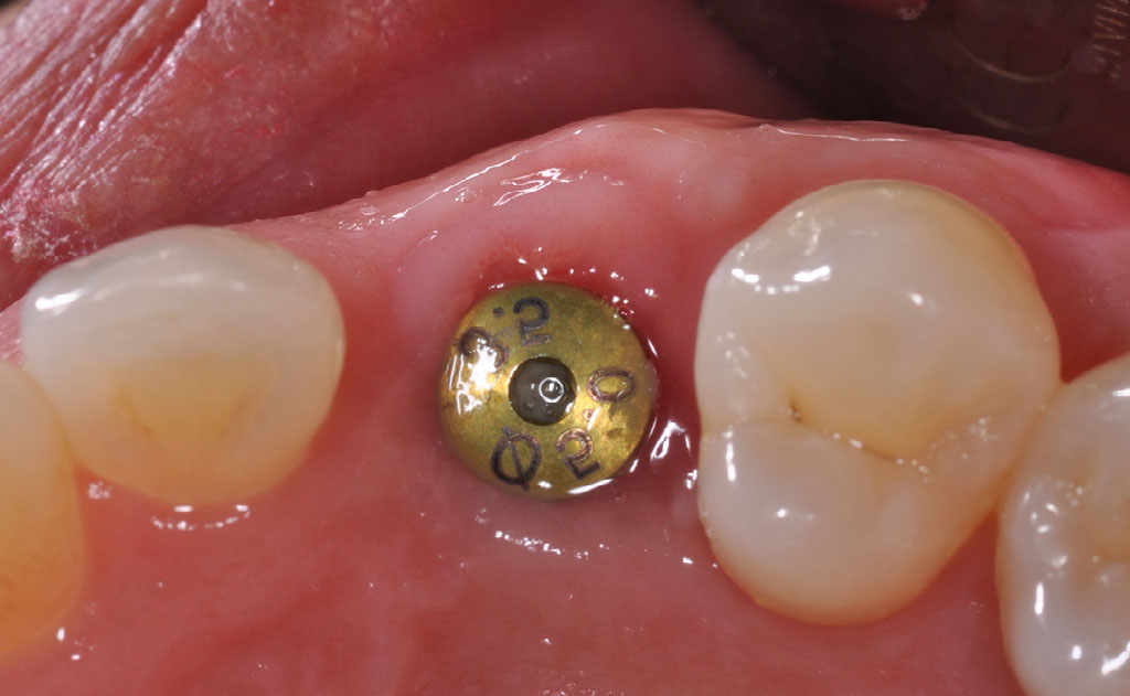
The healing was uneventful
A screw retained restoration was placed
Prosthetics by Dr Ernest Cholakis. Ceramics by Mr. John Verogos, Alpha Ceramics
Replacing the failing tooth with a dental implant
by John Tsourounakis DDS, MS, FRCDC, Diplomate of the American Board of Periodontology
Southwest Specialty Group, Winnipeg, Manitoba